Cardiac Catheterization in Jordan
What is Coronary Angiography? Coronary angiography is a diagnostic procedure that involves imaging the coronary blood vessels in your heart using X-rays. It is performed under local anesthesia, during which a small catheter is inserted through the wrist or groin into the heart, and a special dye (contrast agent) is injected into the blood vessels that supply the heart muscle. Images and digital videos are recorded using special X-ray cameras.
This procedure, also known as cardiac catheterization or coronary angiography, was first performed by Dr. Mason Sones in 1958. It is considered the gold standard for diagnosing coronary artery disease. These images and videos allow the doctor to visualize what is happening inside the blood vessels. Depending on the findings, the doctor may perform procedures to open partially or completely blocked arteries. The procedure also assesses the functionality of the valves between the heart chambers and the pumping strength of the heart muscle.
Understanding the Heart's Artery Anatomy The heart is a muscular organ sustained by millions of blood vessels that supply it with oxygen and nutrients, giving it the energy needed to keep pumping blood. The three main blood vessels of critical importance are:

Left Anterior Descending artery (LAD)
Left Circumflex artery (LCx)
Right Coronary Artery (RCA)
If the heart doesn't receive enough oxygen, it may lead to chest pain or a heart attack.
The Role of the Three Main Coronary Arteries
LAD: Supplies the front and left side of the heart.
LCX: Supplies the left side and sometimes the back of the heart.
RCA: Supplies the right side and bottom of the heart.
Coronary angiography shows how affected or narrowed these arteries are due to atherosclerosis and cholesterol buildup.
What is Coronary Atherosclerosis?

Atherosclerosis is the process through which fatty deposits and cholesterol narrow the coronary artery, depriving the heart muscles of the oxygen and nutrients needed to function properly. Over time, this can weaken or damage the heart. If the plaque ruptures or breaks, the body attempts to repair it by forming a blood clot around it. This new clot can block blood flow in the artery and stop blood flow to the heart, a common cause of heart attacks or angina.
Who Can Benefit from this Procedure?
Patients with angina pectoris.
Patients with symptoms of heart failure or valve disorders.
Those with abnormal heart test results.
People with recurrent fainting episodes.
Patients experiencing chest pain again after a previous catheterization.
Suspected heart attack patients.
Benefits of the Procedure Coronary angiography helps diagnose heart problems and, in many cases, helps the cardiologist treat the issue, relieve symptoms, increase lifespan, and reduce the risk of sudden death, by the grace of God.
Unique advantages of this procedure include:
It is definitive and conclusive.
The results are known immediately.
Treatment with coronary angioplasty (stents) can be performed during the same session once coronary artery disease is confirmed.
It prevents unnecessary tests and medications.
The procedure is used for:
Diagnosing the presence and severity of coronary artery disease and atherosclerosis.
Answering questions such as:
Is there blockage in the heart arteries?
How many blockages are there?
Where are the blockages located?
How severe is the blockage?
What is the best way to treat it?
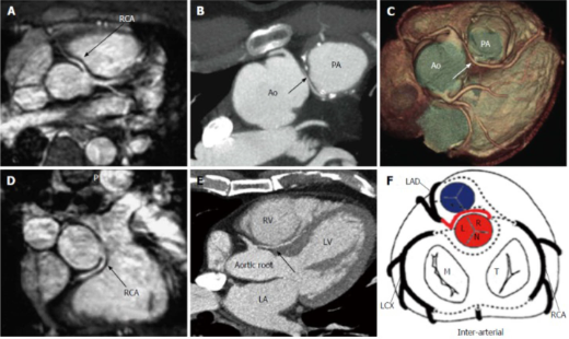
Diagnosing coronary artery abnormalities: Congenital abnormalities in which the origin or path of the coronary artery is anatomically different. In some cases, this can cause a significant reduction in blood flow to the heart muscle, leading to chest pain, irregular heartbeats, or even sudden cardiac death, God forbid.
Assessing the cause and severity of heart failure: A specialized catheter is inserted from your wrist to the left ventricle, and dye is injected to assess the pumping function.
Are There Any Less Invasive Tests for My Condition?

There are other tests available to diagnose coronary artery disease that you may have already undergone. These include:
Electrocardiogram exercise test (treadmill test)
Exercise echocardiography (ESE)
Myocardial perfusion stress imaging
Dobutamine stress perfusion imaging
Multi-slice coronary computed tomography angiography (CTA)
Cardiac positron emission tomography (PET)
These tests provide different types of diagnostic information. However, coronary angiography is generally the most accurate way to confirm coronary artery disease.
Key Facts About Cardiac Catheterization in Jordan
It is a half-day procedure.
You will be asked to fast for 6 hours before the procedure.
The actual coronary angiography takes about 30 to 45 minutes.
If the results are clear, you can go home after 4-6 hours of staying in the day unit.
If stents (mesh) are placed, you may need to stay one night in intensive care and possibly another day in the general ward if necessary.

It's Not Surgery: The procedure is done under local anesthesia. You may be given a mild sedative if you feel anxious.
The catheterization is painless. Any discomfort usually comes from the initial local anesthetic injection on the skin, but the insertion of the diagnostic catheter should not cause more than slight discomfort and no pain.
It is a safe procedure with minimal risks. Coronary angiography is a very common and well-tolerated diagnostic procedure performed more than 2 million times annually worldwide with minimal risk.
Common risks (less than 5%) include bruising and bleeding at the puncture site.
Rare risks (less than 1%) include allergic reactions to the contrast dye.
Extremely rare complications (less than 0.1%) include heart attack, stroke, emergency heart surgery, and death.
How Should I Prepare for the Procedure?
The Day Before the Procedure
Your doctor will explain how the procedure will be done, the risks, benefits, and the expected duration of hospital stay.
Informed consent will be obtained.
Routine blood tests and an electrocardiogram will be performed if not done previously.
Tell your doctor if you have:
Any allergies (especially to iodine, X-ray contrast, or pain medications).
A history of stomach ulcers, recent strokes, or previous bleeding.
Plans for surgery (such as eye, knee, or dental procedures).
A history of kidney problems.
Instructions for medications:
Stop taking metformin (Glucophage) two days before the procedure.
Discontinue blood thinners two days before the procedure under your doctor's guidance. These include:
Warfarin (Coumadin, Marevan)
Dabigatran (Pradaxa)
Apixaban (Eliquis)
Rivaroxaban (Xarelto)
Continue taking aspirin and/or clopidogrel if prescribed by your doctor.
Stay well-hydrated by drinking fluids. This reduces the risk of kidney issues from the contrast dye.
On the Day of the Procedure
Arrival: Go to the admissions office where staff will make you comfortable in your room.
Fasting: You can drink water and have a light breakfast (like cereal/eggs/toast/Milo) up to 4 hours before the procedure. Food will be provided afterward.
Medications: Take all prescribed medications (such as aspirin and/or clopidogrel) with plain water on the morning of the procedure, except diabetes medications (especially metformin). Bring all your medications with you.
Clothing: Wear casual, comfortable clothing, and bring toiletries, an extra set of clothes, and underwear.
Where Will the Catheterization Take Place? The test is typically performed as a day surgery procedure in: The cardiovascular lab in the catheterization department (not in the operating room).
Before the Procedure

You will be asked to empty your bladder before the procedure.
You will change into a hospital gown and be brought to the catheterization department, where your cardiologist and a team of specialized technicians, radiologists, and nurses will be ready to perform the procedure.
The nurse will record your height, weight, and blood pressure and insert an IV cannula into a vein in your arm.
You will lie on a moving X-ray table equipped with X-ray cameras and heart monitors.
Three electrodes will be placed on your chest wall and connected to a heart monitor, which will record the electrical activity of your heart during the test.
You may be given a mild sedative, which will make you feel drowsy.
You will be covered with a sterile drape, and the nurse will clean your wrist or groin (depending on the access site chosen by your cardiologist) with a cold antiseptic solution.
During the Procedure

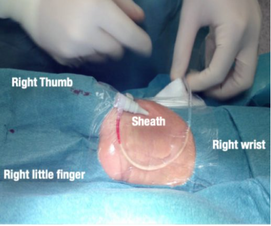
The procedure is divided into two parts. The first part is gaining vascular access, and the second part is obtaining coronary angiography images. A local anesthetic is injected into the skin of the wrist or groin, causing temporary stinging. Once the site is numb, a small plastic sheath (2-3 mm) is inserted into the radial or femoral artery.
This sheath allows a catheter to be inserted into the heart under X-ray guidance. You should feel no more than mild discomfort in your arm during the catheter's movement, and sometimes a warm sensation in your body during the procedure. This usually takes about 5 minutes to complete.
Obtaining Coronary Angiography Images in Jordan
Small amounts of contrast dye will be injected to visualize the coronary arteries and sometimes the left ventricle under X-ray guidance.

The X-ray camera will move around your body to capture images from different angles. This step is generally painless, except for occasional chest warmth.